
We left off last time talking about opaque eye, meaning it in its broadest interpretation, i.e. including all those conditions that induce in the owner the - correct - feeling that their companion's eye has lost transparency.
There are many pathological conditions that can lead to this alteration, so let's start by talking about a particularly serious condition that needs to be addressed promptly.
It will help us to address the Dr. Daniele Santillo, a veterinary ophthalmologist working between Italy and the UK.
How glaucoma is defined in veterinary ophthalmology
 The term glaucoma derives from the Greek 'glaucos' meaning bluish, referring to the colouring that the cornea can take on in the advanced stages of the disease. A 'poetic' term that identifies, however, a devious and painful disease that is an important cause of blindness worldwide in both humans and our animals.
The medical definition of glaucoma has changed over time according to the growing understanding of the disease, however, at least in pets, it continues to be strongly characterised by the increase in intraocular pressure to levels incompatible with normal eye function. Glaucoma leads to irreversible damage to the retina and optic nerve until complete blindness, often accompanied, it should be remembered, by intense pain.
Glaucoma is to all intents and purposes a true ophthalmic emergency and, when there is a suspicion of it, a visit to the veterinary ophthalmologist should be made as soon as possible.
Before we delve into symptoms and treatment, let us briefly understand what 'increased intraocular pressure' means: the inside of the eye is not empty, as there circulates (in the anterior part) a transparent fluid, aqueous humour, which is produced by the ciliary processes (at the back of the iris periphery) and is carried anteriorly up to the anterior chamber (the space between the cornea and the iris) to provide metabolic support for the crystalline lens, the natural lens, and the cornea. From here the aqueous humour exits through the iridocorneal angle (the so-called 'conventional route') and, to a lesser extent, through the iris, ciliary bodies and vitreous (the so-called 'unconventional route') to reach the venous circulation. To summarise and perhaps simplify in the extreme, the ciliary processes can be compared to an always open tap, while the iridocorneal angle can be compared to the drain of the 'eye' hydraulic system.
Intraocular pressure is the result of the balance between production and drainage of aqueous humour, and in a healthy eye it remains relatively constant. If, therefore, there are no situations in which aqueous production increases, the imbalance occurs as a result of reduced drainage.
What causes glaucoma in dogs and cats
Glaucoma in small animals can be classified according to its origin into glaucoma primary o secondary. Alongside this classification, there is also a more 'technical' one, which, based on the state of the drainage angle, defines the open-angle glaucoma o narrow/closed angle. These classifications are not mutually exclusive, but rather complement each other.
Glaucoma Primary has, as the term itself implies, no other cause and therefore arises spontaneously. Its etiopathogenesis is decidedly complex, also involving anatomical alterations of the filtering angle, but is often not completely defined. Primary glaucoma is 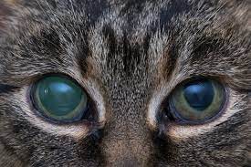 definitely rare in the cat. whereas it is more frequent in the dog, especially in certain breeds that show a recognised predisposition to its development. These are among the most exposed breeds: Beagle, Poodle, Cocker spaniel, Siberian Husky, Samoyed, Great Dane and many others. Primary glaucoma generally affects middle-aged subjects, between 3 and 12 years of age. This clearly tells us that, although probably partly linked to hereditary anatomical alterations, the pathology does not resolve exclusively in them, but has a more complex and, as we said, not always fully clarified origin. Classically, primary glaucoma affects both eyes, although it may not occur simultaneously, with the second eye presenting symptoms up to 12 months after the first. This reiterates the importance of consulting a veterinary ophthalmologist as soon as possible: even if the 'first' eye is now irretrievably blind, much can still be done for the other.
Glaucoma secondarySecondary glaucoma, on the other hand, has no genetic or hereditary basis, but manifests itself as a result of ocular pathologies such as trauma, inflammation (uveitis), dislocation of the crystalline lens or intraocular tumours that mechanically lead to an obstruction of aqueous outflow and/or a closure of the filtering angle. In cats, secondary glaucoma is by far the most common form.
There is also a form of 'congenital' glaucoma, fortunately rare, related to severe eye malformations present from birth and occurring in very young puppies.
What are the symptoms of glaucoma?
Glaucoma is a condition that involves the entire eye and can manifest itself with the following clinical signs:
- Rednesscaused by congestion of the venous vessels of the sclera (the 'white' of the eye around the cornea will appear congested)
-

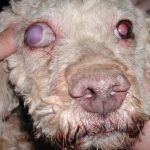
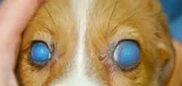
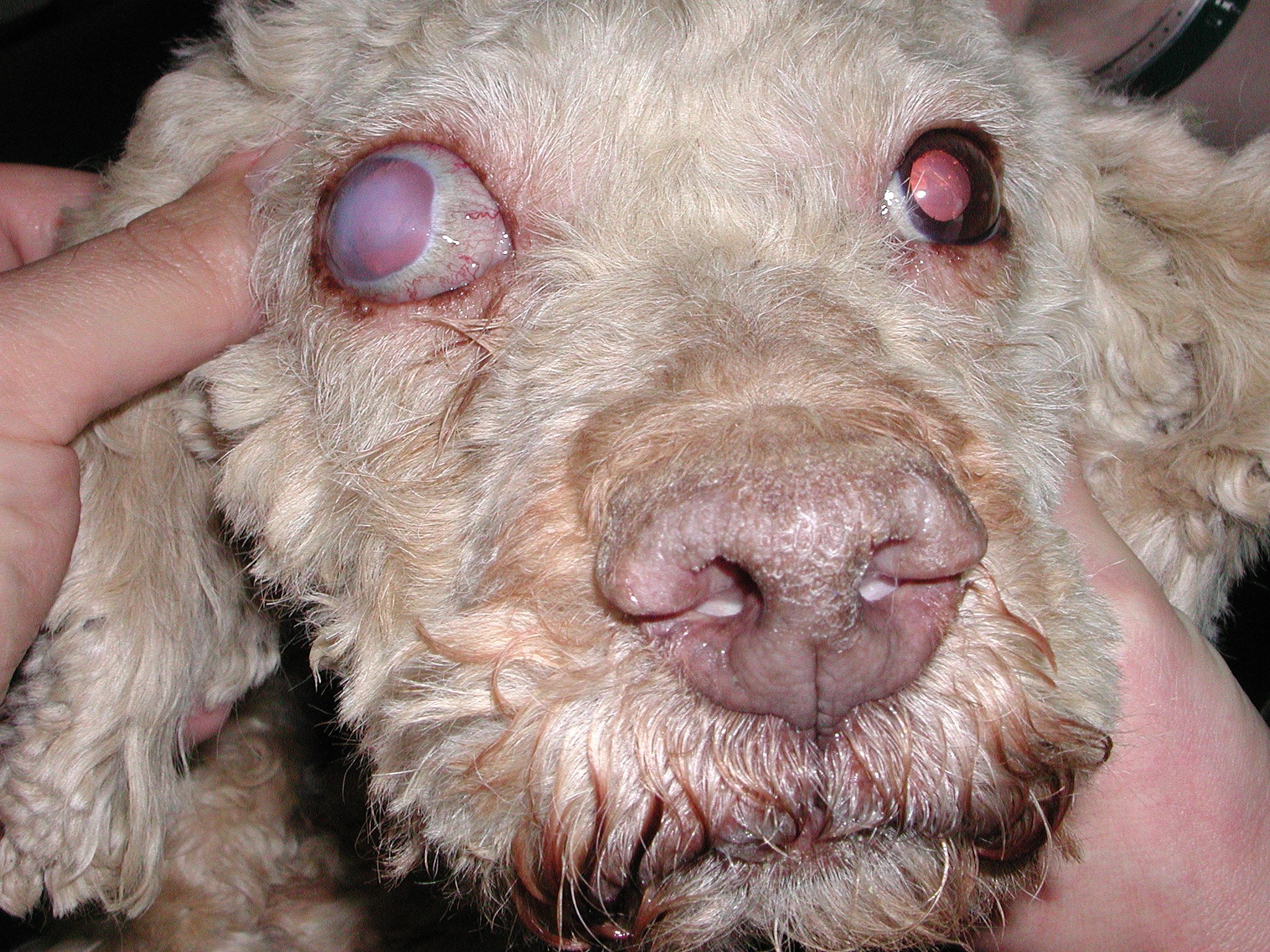
Dog with chronic glaucoma Corneal opacity. Increased intraocular pressure can damage the endothelium, the innermost layer of the cornea (the transparent 'glass' at the front of the eye). As a consequence, oedema can occur, which will cause the cornea to appear bluish, the famous 'glaucous' colour from which the disease derives its name.
- Pain. In dogs and cats it may not be immediately recognisable, but it can be very intense indeed. Human glaucoma patients report sometimes unbearable fierce headaches. Depending on the basic temperament, our animal patient may be extremely depressed or particularly reactive/aggressive, showing particular impatience when touched around the eye. Let us not forget that dogs and cats cannot talk to each other, so let us never underestimate sudden changes in the behaviour of our pet. And when, as we will discuss later, the clinical situation is extreme and requires removal of the eye, let us never forget that owners often report an extreme improvement in the patient's quality of life following surgery to remove the glaucomatous eye (enucleation).
- Visual impairment or sudden blindness. The marked increase in intraocular pressure causes significant suffering of the retina and optic nerve, which reduces visual capacity to the point of complete blindness. Contrary to what one may believe, referring to humans, it is not easy to notice blindness (especially if only of one eye) in our pets. Especially in the domestic environment, dogs and cats are often able to orient themselves very well without the aid of sight. We may perhaps only become aware of the reduced visual ability by changing the position of objects/movements along their usual route,
- Increased eye volume. The strong increase in pressure can cause dilation of the collagen fibres of the sclera and cornea, leading to an increase in the size of the eye (buftalmos). This sign is not early and usually manifests itself in the course of chronic (i.e. very advanced) glaucoma or in very young subjects.
- Pupil dilation. In the early stages it may be moderately dilated, and still respond to light, albeit incompletely. As the disease progresses or in acute cases with very high pressure, the pupil is completely dilated and does not respond to light. The owner will have the impression that he or she can no longer see the colour of the eye or that the eye is 'blacker'.
- Lens dislocation. The crystalline lens may move from its anatomical position (behind the iris, immediately in front of the vitreous) due to the increase in volume of the eye, which causes the fibres holding it in place to break. The opposite can also happen, i.e. the dislocated lens (due to genetic predisposition, trauma, chronic uveitis) can be the cause of glaucoma.
In the following episodes, we will delve into aspects of correct diagnosis and the most appropriate therapy for treating glaucoma in our four-legged friends.
Daniele Santillo

Dr. Daniele Santillo
Med Vet, CertVOphthal, MRCVS
RCVS Advanced Practitioner in Veterinary Ophthalmology
Arezzo - Santillo short CV
In this section:
- Focus on: veterinary ophthalmology
- The opaque eye: not only cataracts...
- Suspected glaucoma: no time to lose!
- Glaucoma: when medical therapy does not work...