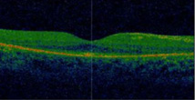
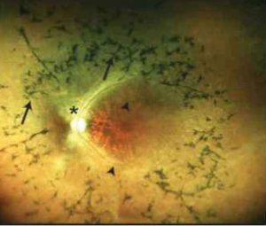
The visual outcome can be predicted on the basis of refractive parameters and OCT data.

The outcome in terms of visual acuity of cataract surgery in patients with retinitis pigmentosa is an extremely delicate and complex issue.
Retinitis Pigmentosa (RP) is a rare genetic disease that results in a progressive degeneration of the photosensitive cells of the retina, causing the gradual loss of night vision and the peripheral visual field and, in its later stages, can even lead to the impairment of central vision and blindness.

A study of RP patients undergoing cataract removal has for the first time identified the condition of the outer limiting membrane and the central macular thickness as parameters for predicting postoperative visual outcomes in RP patients.
109 eyes of 70 MRI patients who underwent a comprehensive eye examination were included in the study, including spectral domain OCT, direct ophthalmoscopy and measurement of best corrected visual acuity (BCVA), before surgery and three months later.
Data were collected on the ellipsoid zone, the second hyper-reflective band of the OCT that corresponds to the anatomical location of the ellipsoid portion of the inner segment photoreceptors and whose distortion reflects changes in the outer segment photoreceptors.
The final results indicated that BCVA was improved in about half of the operated eyes, and the best visual results post-surgery were seen in patients with intact inner limiting membrane, greater macular thickness and higher BCVA levels before surgery.
Source
Mao J, Fang D, Chen Y, et al. Prediction of Visual Acuity After Cataract Surgery Using Optical Coherence Tomography Findings in Eyes With Retinitis Pigmentosa. Ophthalmic Surgery, Lasers and Imaging Retina. 2018;49(8):587-594 https://doi.org/10.3928/23258160-20180803-06
Dr. Carmelo Chines
Direttore responsabile
