


Research at the Luigi Sacco Hospital identifies the eye fundus examination as a useful test in patients with SARS-Cov-2 infection.

Growing and relevant clinical evidence demonstrates the ability of SARS-CoV-2 to attack endothelia and induce major alterations to the blood vessels of many body districts.
This involvement in infection also seems to concern the retinaas described in research carried out by an Italian team and published in the prestigious journal The Lancet.
Retinal findings in patients with COVID-19: Results from the SERPICO-19 study
Invernizzi A, Torre A, Parrulli S, et al.
The Lancet, Vol. 395, Issue 10237,P1610, May 23, 2020
Alessandro Invernizzi and other researchers from the Ophthalmology Unit of the 'Luigi Sacco' Hospital in Milan examined the ocular fundus of 54 COVID-19 patients to identify retinal tissue and vascular changes and compared them with that of 133 subjects who were not infected with SARS-CoV-2.
In COVID-19 patients both the veins and retinal arteries were dilated and the degree of dilatation was positively correlated, to a significant extent, with the degree of severity of the disease and negatively at the onset of symptoms.
This result seems to identify retinal vessel dilation associated with COVID-19, which can be detected even with a single ocular fundus photo, as an important element in assessing the severity and stage of the infection by monitoring the inflammatory response and/or endothelial damage induced by SARS-Cov-2.
Consider that the examination of the ocular fundus practically constitutes a "unicum', which offers the opportunity to analyse vessels 'in vivo', through fundus examination, which is quick, inexpensive and relatively non-invasive.
It is still not entirely clear whether the alteration in the retinal vessels should be interpreted as a specific manifestation of the infection in the ocular district or a consequence of the systemic inflammation induced by COVID-19.
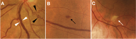
Other abnormalities such as retinal haemorrhages (9.25%), cottony exudates (7.4%) and the development of tortuous vessels (12.9%) were also found in the same patients with retinal microangiopathies.
Subsequent research activity will be aimed at converting this preliminary clinical evidence into the development of a clinical protocol that can be useful for the early detection of patients at risk of developing the severe form of COVID-19, precisely from the early detection of retinal abnormalities.
For a complete overview of this research you can read the full text available online.
Good reading
You might also be interested in:
For the possible correlation between COVID-19 and retinal lesions, we also point out:
COVID-19 and the eyes: scientific studies reveal retinal involvement
Dr. Carmelo Chines
Direttore responsabile