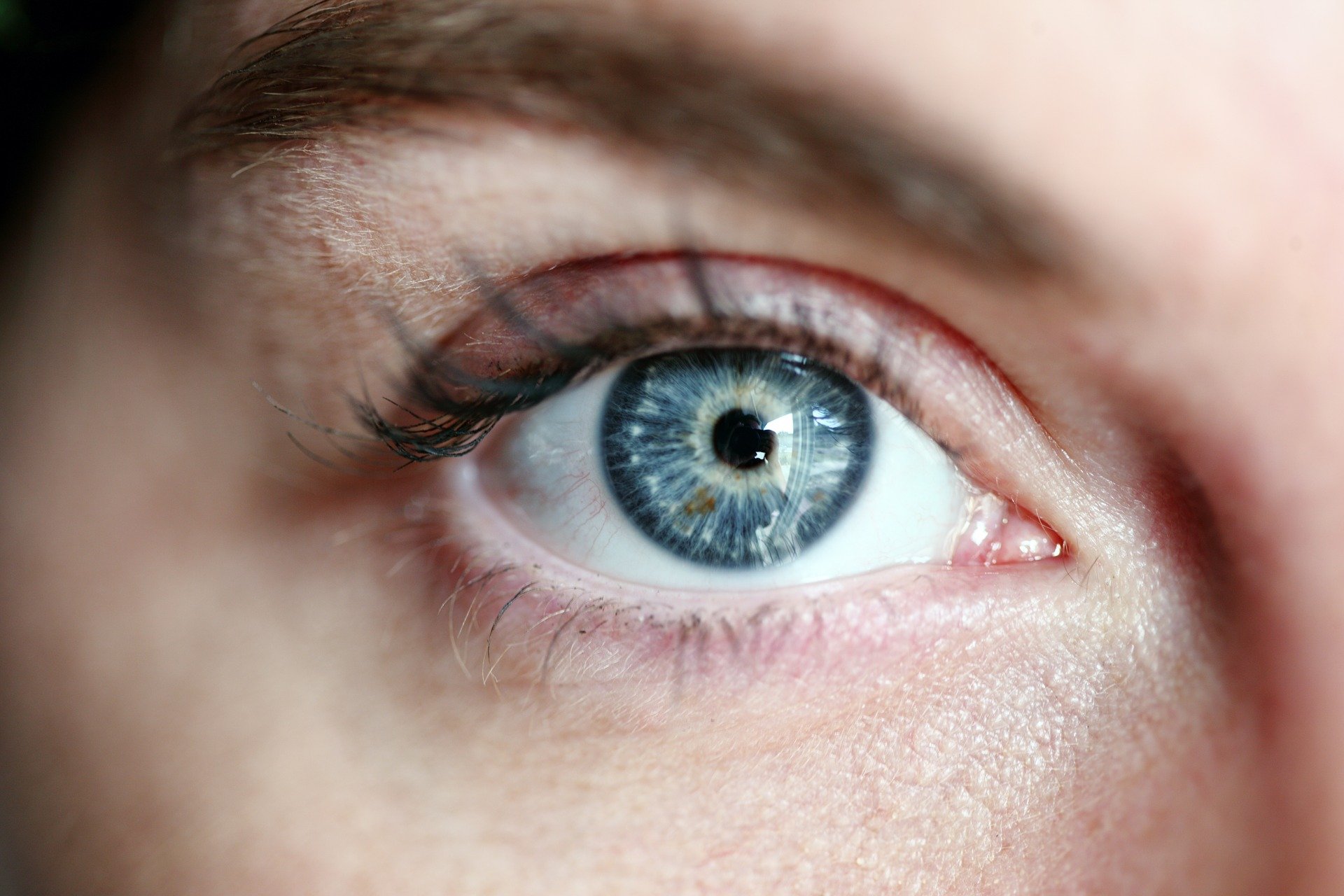
Vitreous detachment, or posterior vitreous detachment, is defined as the separation of the vitreous body also called 'vitreous humour', from the neurosensory layer of the retina.
The vitreous humour is a gelatinous substance between the lens and the retina, composed mainly of water (98%), type 2 collagen and hyaluronic acid. It is surrounded by a translucent membrane, called the hyaloid membrane. In the early period of life, the vitreous is completely attached to the retina.
Some historical background
The vitreous body has, over time, been one of the most studied ocular structures and its actual composition has been the subject of different theories, elaborated according to the knowledge acquired during various historical periods.
More than a century ago, Duke Elder described it as a structure composed of "loose, delicate filaments surrounded by fluid". During the 18th century, this early theory was taken up and modified until four different schools of thought were formed regarding the composition of the vitreous.
The first of these was based on the theory proposed by Demours in 1741, which took the name of 'Honeycomb Theoryin which the idea that the vitreous structure comprised several watertight compartments, or alveoli, containing fluid portions of vitreous was expounded.
In 1780, Zinn hypothesised that the vitreous could be organised in lamellar, concentric layers, similar to those of the onion. This second theory is known as 'Lamellar Theory' and during this historical period, this theory was supported by numerous other scholars of human anatomy.
The "Radial Sectors Theory, elaborated by Hannover in 1845, illustrated how the vitreous structure could be composed of sectors oriented radially around the Cloquet Canal, which was thus placed in the centre (almost simulating the structure of an orange).
The last of these four theories was taken up in 1848 by Bowman and was called "Fibrillar Theory. It was based on his studies using a microscope capable of highlighting the nucleus of the small fibrils that formed the bundles of the vitreous structure.
The changes related to the vitreous gel, with the consequent loss of its transparency, led Szent-Gyorgito to assume that the structure might undergo age-related changes.
For the first time, the fact that thickenings and, therefore, opacities, the so-called ''opacities'', were created in the vitreous.floaters' o 'Flying flies'capable of disturbing vision.
The vitreous body: what is it?
We now know that the vitreous body, or vitreous gel, consists of transparent, gelatinous connective tissue that is neither vascularised nor innervated. Weighing about 4 grams, it makes up 2/3 of the entire volume of the eyeball and because of its size contributes to the shape and substance of the eye.
Due to its high viscosity, the vitreous acts as a shock absorber for possible bulbar trauma, protecting the most delicate ocular structures. In addition to these numerous properties, its elasticity allows antero-posterior displacements of the crystalline lens, helping the ciliary muscle in its accommodative activity.
Composition
The vitreal gel consists of 99% water and the remaining 1% collagen fibrils and hyaluronate, which form the 'scaffold'. The network of collagen fibrils forms a solid structure that is 'inflated' by the hydrophilic hyaluronate, creating the actual vitreal structure. The relationship between these two elements is crucial to maintaining the transparency and structure of the vitreous, as if the proportions between the two were to change (as happens, for example, during ageing) the vitreous would become more liquid and less 'gel-like'.
Fibrils consist of type I and type II collagen with a triple helix structure.
Vitreous structure
The study of vitreous anatomy has always been difficult for two main reasons:
- having to study a completely transparent tissue, a condition that makes all attempts to define the morphology of the vitreous very difficult.
- the techniques used so far to define vitreous structures have been thwarted by the artefacts induced by tissue fixatives, which cause glycosaminoglycans to precipitate.
The vitreous body is to be understood as a dioptric and nutritive structure, as well as morphostatic. It is surrounded by the hyaloid membrane and relates posteriorly with the inner limiting membrane of the retina and anteriorly with the fibres of Zinn's zonula, the ciliary bodies and the posterior face of the lens where it forms a cavity called the patellar fossa.
In some places, the vitreous forms physiological adhesions, especially at the level of the posterior capsule of the crystalline lens through the ligament of Wieger, particularly at the level of the optic papilla (Martegiani's area), and also at the level of the ora serrata where adhesion is particularly high. In adolescence, strong adhesions are present, also at the macular level, but these diminish as the years go by.
The development of the vitreous
Vitreous development begins during the sixth week of embryonic life and, although the origin of cell development is still not entirely clear, it is assumed that vitreous synthesis is due to hyalocytes, retinal cells and ciliary body cells.
During embryogenesis, the vitreous is invaded by blood vessels, which provide nourishment to the developing anterior segment of the eye. Over time, these vessels undergo regression until they disappear completely, leaving the vitreous body transparent.
Its volume increases threefold from birth to adulthood; however, it should be noted that 70% of this increase occurs by the age of four. Complete development is generally reached in the adolescent period, between 15 and 18 years of age.
The stages of development
The formation of the vitreous gel consists of three different moments that contribute to the composition of the final vitreous:
- Primary vitreous: mesodermal tissue surrounds the lenticular vesicle, while in its inner thickness, a vascular web from the hyaloid artery develops.
- Secondary vitreous: It usually forms around the second month of intrauterine life, through a process of transformation of mesenchymal tissue with typically avascular neuroectodermal tissue.
- Tertiary vitreous: characterised by the disappearance of the hyaloid vascular tree and consequently the appearance of the Cloquet Canal. This portion of the vitreous represents a virtual cavity and is optically empty, composed of the primordial mesenchymal tissue now replaced by the definitive vitreous.
Anatomy of the vitreous
Anatomically speaking, the vitreous body is usually subdivided into three different well-defined zones that are called: Central Vitreous, Vitreous Base and Vitreous Cortex.
The Central Vitreous is, as its name suggests, the portion in the centre of the entire vitreous gel which contains within it the Cloquet Canal, which is a remnant of the primary vitreous vascular system during embryonic development. The central vitreous has a low concentration of collagen fibres running antero-posteriorly.
The Vitreous Base is located at the ora serrata, the transition portion between the retinal tissue and the ciliary body. In this anatomical area, the vitreous is characterised by the presence of dense bundles of collagen fibres strongly adhered to the retina, probably due to their 'fusion' with the inner limiting membrane.
Vitreous cortex: the central vitreous gel is in turn surrounded by the vitreous cortex, which has a different orientation of collagen fibres than the central vitreous. The cortex is commonly subdivided into:
– Anterior vitreous cortex: covers the posterior surface of the lens at the patellar fossa.
– Posterior vitreous cortex: this vitreal portion adheres to the innermost retinal surface behind the posterior edge of the vitreous base.

Vitreous detachment
The causes
The main cause of posterior vitreous detachment is advancing age. Normally, in a young, healthy individual, the vitreous is adhered to the inner limiting membrane, which defines the transition from the vitreous body to the retina.
With age, the vitreous loses its gelatinous consistency and tends to degenerate. This process begins with the liquefaction phase of the vitreous, called synchysis, and continues with the syneresisi.e., the aggregation of collagen fibrils, which leads to the collapse of the vitreous. This event produces thick bundles of collagen fibrils that float in the vitreous and give rise to vitreous mobile bodies, the 'Flying flies' or in scientific language 'myodesopsies'.
Degeneration of the vitreous also causes weakening of the vitreoretinal adhesion, which is precisely the cause of posterior vitreous detachment.
This is a spontaneous process, but can be brought about by events such as cataract surgery, trauma, uveitis, panretinal photocoagulation and laser capsulotomy.
Risk factors
The most important risk factors include:
- - age: the incidence of posterior vitreous detachment after the age of 50 is 53% and between the ages of 66 and 86 is 66%;
- - female sex: the progression of a posterior vitreous detachment is faster in women than in men aged 60 years or older;
- - myopia;
- - underlying diseases, such as retinitis pigmentosa;
- - menopause: post-menopausal female patients may be more prone to develop this condition, due to oestrogen deficiency;
- - vitamin B6, this vitamin has an anti-estrogenic effect, so a higher intake of vitamin B6 may increase the incidence of posterior vitreous detachment in women;
- - long-lasting inflammation, resulting in cell proliferation which, in the long run, can cause fibrosis. Vitreous fibrosis causes, in turn, traction on the retina, which can lead to posterior vitreous detachment or retinal rupture;
- - trauma: posterior vitreous detachment can occur as a result of penetrating trauma, i.e. an eye injury caused by sharp or pointed objects;
- - some eye surgery procedures.
Symptoms
In most individuals, the early stages of posterior vitreous detachment are asymptomatic and are not detected clinically until the separation of the vitreous from the margins of the optic disc produces symptoms.
The main symptoms are flashes of light e floaters (myodesopsies). 67% of patients complain of blurred vision, which may occur due to haemorrhage of the vitreous from retinal breaks or mobile bodies crowding the visual field.
Diagnosis
A complete retinal examination must be performed to confirm the diagnosis. The key diagnostic procedures in the evaluation of acute posterior vitreous detachment are binocular indirect ophthalmoscopy and three-mirror lens biomicroscopy. Imaging-based diagnosis traditionally involves the use of dynamic B-scan ultrasound. More recently, optical coherence tomography has been added to the most commonly used imaging diagnostic techniques.
Complications and prognosis
The most important complications of posterior vitreous detachment are:
- - retinal ruptures
- - retinal detachment
- - vitreal haemorrhage
- - retinal haemorrhage
- - cystoid macular oedema
- - macular hole
How to treat vitreous detachment
If no complications are present, posterior vitreous detachment usually has a good visual prognosis and does not require any treatment.
In the case of an abnormal detachment, which leads to complications, early diagnosis and timely recourse to the most appropriate treatment for each pathological condition (retinal rupture or detachment, haemorrhage, macular hole, etc.) are essential.
How to prevent vitreous detachment
There is no protocol or rules to prevent vitreous detachment, as it is a phenomenon linked to the natural ageing process of the eye.
Practical advice
To try to prevent or slow down vitreous detachment, it is important to follow a few simple recommendations:
- Drink plenty of water, at least 1.5-2 litres a day
- Consuming foods rich in antioxidants and omega 3, such as fruit, vegetables, fish and nuts
- Avoiding eye trauma
- Have regular eye check-ups and promptly report any symptoms to your doctor
The warning signs
Blurred vision, flashes of light or moving bodies
- Faryal Ahmed, Koushik Tripathy, Posterior Vitreous Detachment, StatPearls [Internet]. Treasure Island (FL): StatPearls Publishing; 2021 Jan. 2021 Feb 14.
- Huang LC, Yee KMP, Wa CA, Nguyen JN, Sadun AA, Sebag J. Vitreous floaters and vision: current concepts and management paradigms. In: Sebag J, ed. Vitreous - in Health and Disease. New York, NY: Springer; 2014:771-788
