


L'diabetic macular oedema (EMD) is the main cause of visual impairment in individuals with diabetic retinopathy (DR). Its incidence increases predominantly with duration of diabetes, as well as the presence of other conditions such as hyperglycaemia, nephropathy or hyperlipidaemia (high blood lipids).
In order to reduce the likelihood of the onset and progression of EMD, the EMD sufferer must monitor certain parameters, such as:
-glycaemia;
-blood pressure;
-triglyceride and cholesterol levels in the blood.
Diagnosis
The diagnosis of diabetic macular oedema is made by careful examination of the ocular fundus, examinations may include:
-the biomicroscopy, examination by means of a slit lamp used to carefully assess both the bulb and the ocular adnexa;
-the fluoroangiography, which highlights any retinal lesions;
-the tomography at optical coherence (OCT) which is necessary for the quantification of retinal thickness and oedema.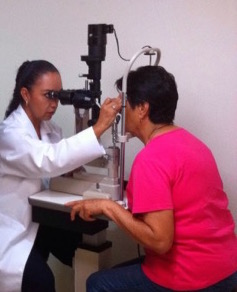
Therapeutic Strategies
Laser treatment
Laser photocoagulation
The traditional treatment of EMD, introduced as early as the 1980s, was carried out with laser photocoagulation (focal or grid treatment). Its effectiveness in preventing vision reduction and loss has been proven by data from two large studies: the Diabetic Retinopathy Study (DRS) and theEarly Treatment Diabetic Retinopathy Study (ETDRS). The laser photocoagulation is used to treat microaneurysms and areas of focal loss that do not involve the centre of the retina. The results of the ETDRS study have, in fact, demonstrated a reduction in the risk of vision loss in the 50% eyes under study following laser treatment. This type of treatment, despite being very effective, could be responsible for certain complications such as comparation of scars within the retina or paracentral scotoma.
Diode laser
Diode laser photocoagulation is a treatment that uses a specific wavelength that is absorbed by the outer layers of the retina, thus reducing the risk of possible complications such as scotoma.
Intravitreal therapy
Anti-VEGF
Anti-VEGF drug therapy is able to inhibit the production of the vascular endothelial growth factor (VEGF) responsible for the uncontrolled formation of new blood vessels. Despite the proven efficacy of this treatment, these molecules have a time-limited therapeutic effect that may result in the EMD sufferer having to undergo several intravitreal injections to ensure long-lasting efficacy.
Steroids
Steroid drugs are anti-inflammatory molecules used to treat diabetic macular oedema because they can inhibit the processes underlying the onset of EMD, including inflammatory ones; are in fact able to:
-stabilise certain endothelium cells;
-inhibit leucocyte migration, as well as the synthesis of vascular growth factor, cytokines and prostaglandins.
Corticosteroid therapy, such as fluocinolone acetonide, is recommended for patients with EMD who are not sufficiently responsive to anti-VEGF drugs or who cannot be treated with these molecules and who also meet the requirements for steroid treatment (aphakia and absence of ocular hypertension or glaucoma)
Surgical treatment
Surgery involves the removal of the vitreous body (vitrectomy). This is performed only in more severe cases characterised by the presence of ocular haemorrhages or retinal complications.
Sources:
Guidelines for the screening, diagnosis and treatment of diabetic retinopathy in Italy, Working Group on Ocular Complications of Diabetes, Italian Society of Diabetology, 2015
Dr. Carmelo Chines
Direttore responsabile