


The myasthenia gravis (MG) is a chronic autoimmune disease, which involves the neuromuscular junction and which in the majority of patients, approximately 90% of cases, results in ocular involvement that manifests itself with clinical pictures of varying severity and with symptoms of blurred vision and problems with balance and unsteady gait.
Myasthenia gravis is a rare diseasewith an estimated prevalence of 1/5,000 and an incidence of 1/250,000-1/33,000 in Europe. It affects both sexes, with a difference in the time of onset, which in females occurs predominantly before the age of 40 (early onset), while in males after the age of 50 (late onset).
The pathogenesis of myasthenia gravis is autoimmune and is mediated by pathogenic antibodies against specific target antigens of the post-synaptic membrane, including antibodies against the acetylcholine receptor (AChR), which are found in 85% of cases of generalised myasthenia.
The prevailing symptom is fluctuating muscle weakness and, in particular, fatigability of the eye and bulbar, skeletal and/or respiratory muscles due to involvement of the striated muscles. This weakness is caused precisely by the attack by autoantibodies directed against neuromuscular junction proteins. The clinical severity of myasthenia gravis can range from mild ocular symptoms to frequent respiratory crises.
Among the striated muscles most frequently affected at the onset of the disease are: the extraocular muscles (in 85-95% of cases), the bulbar musculature (with dysphagia and dyspnoea in 20% of cases) and the facial and oropharyngeal musculature (with hypophonia, rhinolalia, dysarthria, masticatory fatigability and facial weakness in 15% of cases), the proximal muscles of the limbs (in 8-20% of cases)
When the muscle weakness is spread to various districts, we speak of generalised myasthenia gravis, while when only the district of the extrinsic 0ìocular musculature is affected, it is called ocular myasthenia gravis.
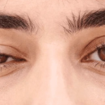
The most frequent ocular symptom is the ptosiswhich may be unilateral or bilateral, associated with diplopia (double vision). Moreover, one of the most frequently observed clinical signs is the different set-up of the two eyelids: the one less affected by ptosis is usually hyper-retracted, while the contralateral one is completely drooping, with passive elevation.
The factor that triggers the autoimmune response remains unknown, but a frequent association with pathologies of the thymus, the gland of the immune system, has been observed in Ab AchR+ forms (60-70% hyperplasia, 10-12% thymoma) and a clinical improvement after thymectomy is noted, which has led to speculation of a central role of the thymus in the production of the antibodies responsible for the disease.
Diagnosis
In addition to the objective clinical examination, some specific tests are used:
- Edrophonium chloride test, which is injected to see if muscle strength improves and if this happens, it could be an indication of myasthenia gravis.
- Try the ice pack, which is placed in the periocular area for two minutes to examine whether the cold sensation affects ocular motility.
- Routine blood tests + anti-AChR+, anti-MUSK, anti-LRP4 autoantibody tests
- Repetitive Nerve Stimulation (NR): small electrical impulses are sent through electrodes placed in the muscles to examine the response of the nerves.
- CT scan or MRI to rule out a tumour in the thymus area.
- Pulmonary function test to check respiratory capacity
Treatment
Given the prevalent ocular onset of this disease, it is of great importance that ophthalmologists are able to make an early diagnosis, applying the criteria for a differential diagnosis, and are up-to-date on currently available and developing therapies to be used, often in coordination with the neuroimmunology specialist.
Myasthenia gravis, in fact, is a condition that can be cured, even if not cured, provided it is diagnosed early and given appropriate treatment.
Traditional therapy involves symptomatic treatment with cholinesterase inhibitors, long-term administration of steroid drugs and immunomodulation strategies, which may also involve surgical removal of the thymus.
However, the burden these therapies place on the patient in terms of long-term side effects is very significant and new therapeutic alternatives are currently being developed, often as a result of biomolecular research.
Among the therapies currently undergoing clinical trials (which we invite you to learn more about on the page Experimental Treatments for Myasthenia Gravis | Myasthenia Gravis News we report a range of antibody-based therapies, such as Batoclimab, DNTH103, Inebilizumab, and a range of T-cell-based therapies, such as CABA-201, Descartes-08 KYV-101 and MuSK-CAART
It is also possible to suggest lifestyle modifications to the patient, which can be supportive of drug treatment, such as:
- frequent breaks while reading or working at the computer, to avoid eye overexertion
- adequate lighting conditions in the living and working environment
- have a comfortable chair and table to reduce or prevent muscular stress on the entire body, which affects the eyes
- in the case of ptosis, use special scleral contact lenses to support the drooping eyelid
- in case of diplopia, use prismatic lenses that help realign the image in the brain.
On the subject of rare diseases, we also propose:
- Hereditary maculopathies: genes & C - Oculista Italiano
- Stargardt's disease: new hope for patients - Oculista Italiano
- Ocular syphilis and early diagnosis - Oculista Italiano
- Cogan's syndrome: rare but treatable - Oculista Italiano
- Jacob, S. Treating myasthenia gravis beyond the eye clinic. Eye38, 2422-2436 (2024). https://doi.org/10.1038/s41433-024-03133-x
- Iorio, R. Myasthenia gravis: the changing treatment landscape in the era of molecular therapies. Nat Rev Neurol20, 84-98 (2024). https://doi.org/10.1038/s41582-023-00916-w
- Myasthenia Gravis News Home