


In a recent study, published in the journal Stem Cellmini-brains, grown in the laboratory from stem cellsThey spontaneously developed rudimentary eye-like structures. On these tiny human-derived brain organoids, cultured in vitroScientists at the University Hospital Düsseldorf, Germany, have observed the bilateral growth of two symmetrical eye cups, mirroring the development of eye structures in human embryos. This incredible result may improve our understanding of the process of differentiation and development of the eyes, as well as the mechanisms behind many eye diseases.
"Our work highlights the remarkable ability of brain organoids to generate primitive light-sensitive sensory structures that house cell types similar to those found in the human body." - said neuroscientist Jay Gopalakrishnan from the University Hospital of Düsseldorf -"These mini-brains will be useful in the study of brain-eye interactions during embryo development. They will also provide a model for studying congenital retinal disorders and generate patient-specific retinal cells for personalised drug testing and transplantation'.
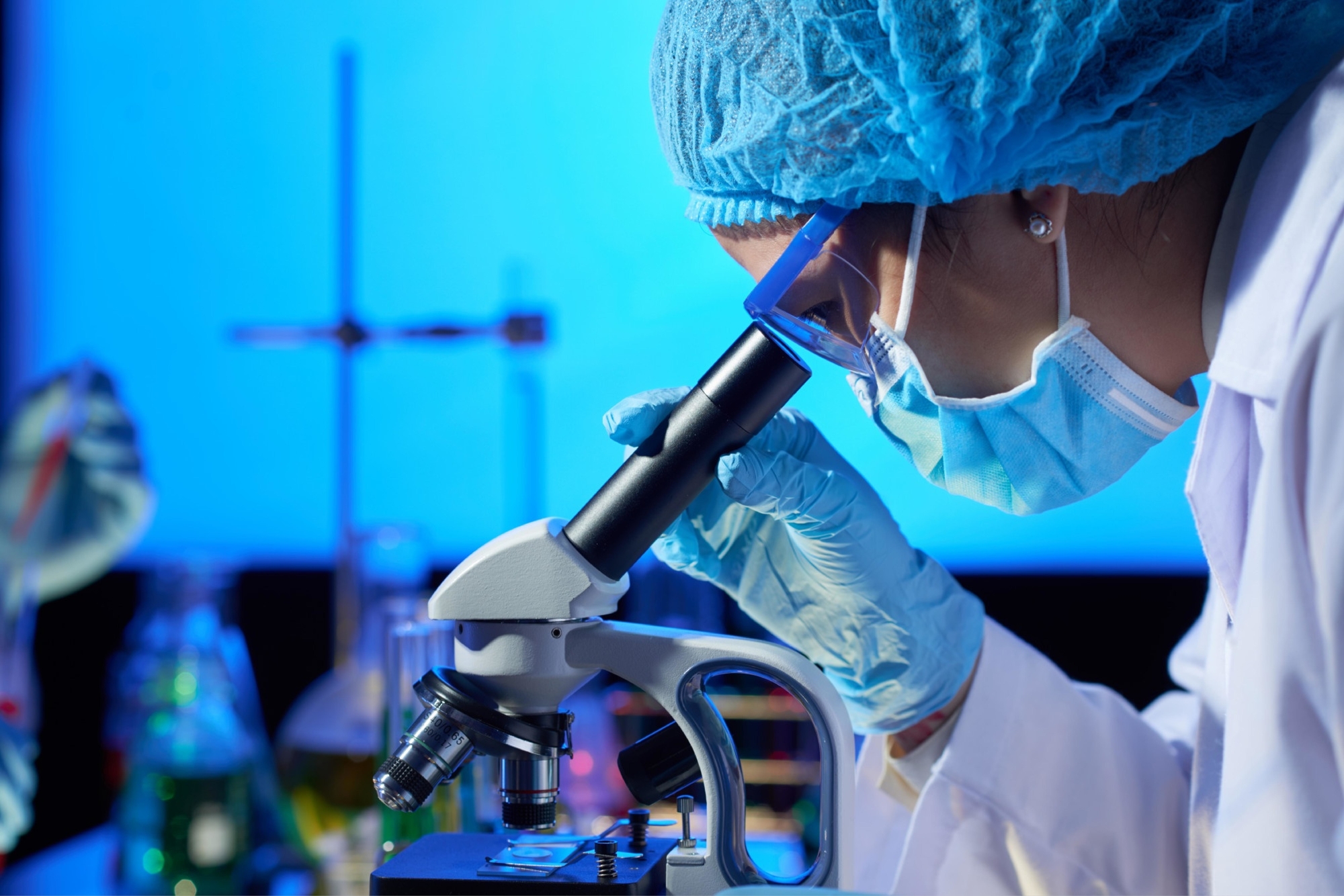
Brain organoids are clearly not real brains. Instead, they are small three-dimensional structures grown from induced pluripotent stem cells - cells harvested from adult humans and reprogrammed into stem cells - that have the potential to differentiate into many types of tissue.
In this specific case, stem cells were pushed to grow and differentiate into brain tissue. These 'mini brains' are used for research purposes where the use of real living brains would be impossible, or at least ethically complicated. They are useful, for example, to test responses to drugs or to observe cell development under certain adverse conditions.
In previous research, other scientists had used embryonic stem cells to cultivate optic cups, the structures from which eyeballs originate during embryonic development. And other groups had developed similar optical structures from induced pluripotent stem cells. Rather than cultivating these structures directly, Gopalakrishnan's team aimed to grow them as an integral part of brain organoids, with the advantage of being able to observe how the two types of tissue could grow together and interact.
"Eye development is a complex process and understanding it could provide a better understanding of the molecular basis of retinal diseases." - the researchers wrote in their article - "Therefore, it is crucial to study the optic vesicles, present early in the embryonic development of the eye, the proximal end of which is attached to the forebrain, which is essential for the correct formation of ocular structures.
In previous work, during the development of organoids, the scientists had already observed the presence of retinal cells, but these had not developed optical structures. Therefore, the team changed the experimental protocol and, through the addition of retinol acetate to the culture medium, aided ocular differentiation. Under these conditions, mini-brains formed optical cups as early as 30 days of development, with the structures clearly visible at 50 days, which is consistent with the timing of eye development in the human embryo, meaning that these organoids could be useful for studying the intricacies of this process.
There are also other implications: the optical cups obtained in this study contained different types of retinal cells, organised in neural networks, that responded to light and even contained a lens and corneal tissue. Finally, the structures showed retinal connectivity with regions of brain tissue. 'In the mammalian brain, the nerve fibres of the retinal ganglion cells extend to connect with their brain targets, something that has never before been shown in an in vitro system', said Gopalakrishnan.
These results proved to be reproducible: of the 314 brain organoids cultured by the team, 73% developed optical cups. The research team hopes to develop new strategies to keep these structures in culture on longer time scales, in order to carry out more in-depth research.
"Brain organoids containing optic vesicles that display highly specialised neuronal cell types can be developed, paving the way for the generation of customised organoids and retinal epithelium sheets for transplantation." - the scientists wrote in their article - "We also believe that these next-generation organoids will be useful for studying retinopathies that arise in the early stages of neurodevelopment.
Bibliography
Dr. Carmelo Chines
Direttore responsabile