


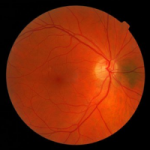
The macular hole (MH, Macular Hole) is a full-thickness retinal defect in the foveolar area leading to impairment of central vision.
The macular hole idiopathic senile occurs in individuals above of 60 years of agewith an estimated incidence of 1 in 5000 subjectsof which, women are more affected than men with a 2:1 ratio. Although spontaneous closure of the macular hole has been reported in some cases and treatment with intravitreal ocriplasmin has shown good results, the surgical approach remains the main one and with the highest anatomical success rate (90%) in the case of MH patients. Most studies conducted to date have reported follow-up 1 year after eyehole surgery and some with longer-term information have shown that Best Corrected Visual Acuity (BCVA) can continue to improve for several years after surgery. However, analyses have never gone beyond five years of follow-up.
Anatomical and visual outcomes following macular hole surgery
A very recent study, published in Ophthalmology Retina, instead assessed the Anatomical and visual outcomes, at more than 5 years of follow-up, in patients with idiopathic macular hole undergoing Vitrectomy Pars Plana (PPV, pars plana vitrectomy) with internal limiting membrane peeling (ILM, internal limiting membrane). This study is the first to report the results of follow up after macular hole surgery at more than 5 years, assessing at the same time possible prognostic factors that may influence postoperative visual outcomes.
The main parameter of success evaluated by the authors was the highest visual acuity correct (BCVA) recorded before surgery and, if available, after 1, 2, 3, 5, 8 and 10 years. This was subsequently correlated with parameters assessed by spectral-domain optical coherent radiation tomography (OCT)such as the restoration of the postoperative integrity of the ellipsoid zone (EZ) and external limiting membrane (ELM) and the presence of cystoid spaces.
The study evaluated 87 eyes from 80 patients with an average age, at the time of surgery, of 68.9± 7.03 years; the follow-up postoperative mean was 9.6±4.3 years. The maximum corrected visual acuity (BCVA) average, recorded before surgery, was 0.20±0.14D, and showed a significant improvement (P < 0.05) in all follow-up. Improvement in BCVA remained stable over 10 years after macular hole surgery. The success in closing the macular hole was achieved in 94% eyeswhile a reopening was registered in 8.0% of the cases. 13% of the eyes underwent a second operation due to persistence or reopening of the macular hole. Indocyanine green staining (ICG) was used in 22 eyes (25.2%). The postoperative integrity of the ellipsoid zone (EZ) was restored in 52 eyes (60%), the integrity of the outer limiting membrane (ELM) in 54 eyes (62%) and cystoid spaces of varying severity were evident in 28 eyes (32%).
The study concludes that the improvement in maximum visual acuity after PPV for macular hole is continuous in the first three years of postoperative care and is maintained in subsequent years in a substantial percentage of patients. In addition, the maximum visual acuity achieved correlated with better preoperative BCVA and better morphological characteristics of the outer retinal layer, assessed by OCT.
The study results support and extend previously published evidence of a positive effect of internal limiting membrane peeling in achieving higher rates of macular hole closure and lower rates of macular hole complications and reopening.
Source:
Elhusseiny et al. Long-Term Outcomes after Macular Hole Surgery. Ophthalmol Retina, 2019.
Dr. Carmelo Chines
Direttore responsabile