The human ocular microbiota
In the human eye coexist numerous bacterial microhabitats, whose composition reflects levels of exposure to the external environment. Studies that have dealt with this topic have shown that the gender most represented on the ocular surface is the Corynebacterium, followed by Staphylococcus, Streptococcus, Acinetobacter e Pseudomonas.
Knowing, however, the distribution of microorganisms in specific ocular areas would be an additional weapon to address eye diseases such as contact lens inflammation/infection, blepharitis, Meibomian gland dysfunction, exogenous endophthalmitis in a targeted manner and would provide a better understanding of ocular surface disorders with an inflammatory component (including episcleritis, chronic follicular conjunctivitis, dry eye syndrome).
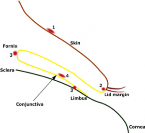
This study, recently published in The Ocular Surfaceanalysed the structure and distribution of bacterial communities in the different microhabitats of the eye human, to understand their similarities and differences. The four eye sites analysed by the team were: eyelid margin tissue of patients with eyelid abnormalities, conjunctival tissue of fornices and limbus of patients with pterygium, ocular surface swabs (conjunctival) and swabs of the facial skin.
In total, the researchers identified 2465 taxonomic units zero-distance operating units (zOTUs), comprising 16 phyla and 338 genera. The study revealed no differences in the zOTU richness of the bacterial communities with respect to gender, but differences were significant by age and between the four regions. The richest area with 188 ± 79 zOTU was the skin, while the poorest with 49 ± 7 zOTU was the conjunctiva.
Ocular bacterial distribution
The study proposed a classification of ocular bacterial distribution into three groups:
- Group 1consisting of zOTUs that are resident in the skin and eyelid margin and move from there to the ocular surface, as indicated by a low relative abundance at this site. The genera belonging to this group are Corynebacterium e Staphylococcus. The former was constantly present on the outer surface of the skin, and its relative abundance progressively decreased as it moved from the skin (12.2%), to the eyelid margin (7.6%), to the ocular surface (4.0%), but was rarely present in conjunctival tissue samples. Similarly, Staphylococcus, a common resident of the skin, was abundant on the skin surface (15.1%), decreased in the eyelid margin (1.9%) and was present in relatively low amounts on the ocular surface (3.1%).
- Group 2consisting of zOTU localised mainly on the ocular surface and detected at low levels on the skin, eyelid margin and conjunctiva. Members of this group could be acquired from air or water and may be able to survive on the ocular surface, but not in other regions of the eye. Acinetobacter, for example, it was isolated in relatively greater abundance on the ocular surface (12.3%) than on the skin (1.0%) and eyelid margin (0.5%)
By Jerome Ozkan et al, 2019
- Group 3, consisting of zOTU present in the conjunctiva and inside the eyelid margin and found inconsistently and in relatively low abundance on the skin and ocular surface. To this group belongs the genus Pseudomonas. Not all members of this group, however, showed the same distribution; some were consistently present in the conjunctiva and eyelid margin, others were more specific to the conjunctiva, and one was specific to the eyelid margin.
The study shows that the micro-organisms in and around the eye are clearly spatially organised, and this biogeography is representative of a process of microbial dispersion and environmental selection.
Source:
Biogeography of the human ocular microbiota. Jerome Ozkan et al. The Ocular Surface, 2019
Dr. Carmelo Chines
Direttore responsabile

