


With the approach of summer, the dangers of transmitting viral infections through mosquito bites are reappearing.
The summer and, in general, the hot-humid climate due to global warming are at the root, even in our country, of a significant increase in the health risk from mosquitoes, especially certain particularly aggressive or exotic species.
As is well known, mosquitoes can be vectors of viral infections such as Dengue, yellow fever, Chikungunya and Zika. We have dedicated an in-depth study published in our paediatric ophthalmology section to the alterations in visual function caused by the latter virus. Zika virus and visual impairment - Oculista Italiano
In this article, we would like to present a case report on theWest Nile Virus infection (WNV), carried out by an Italian research team from the University of Bologna, led by Prof. Nicola Valsecchi
West Nile Disease (WND) is a zoonosis of viral aetiology, transmitted by mosquitoes, that causes forms of meningo-encephalitis in birds, both wild and domestic, equids and humans.
In the case report we present, however, the only manifestation of acute West Nile virus infection was a bilateral multifocal chorioretinitis.
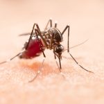
Mosquitoes: viral vectors
WNV is transmitted by different genera and species of mosquitoes. The most commonly detected vectors in outbreaks belong to the genus Culex e Aedes spp. In Italy Cx. pipiens s.l. appears to be the species most involved in the circulation of the infection. Generally, propagation by mosquitoes is reduced during the colder months, but it has nevertheless been shown that the virus is able to survive during this period in infected mosquitoes that overwinter in warmer areas.
Symptoms and mortality
In humans, approximately 80% of cases of WNV infection develop asymptomatically. The infection generally has a seasonal pattern, with most cases detected between July and October, peaking in late August.
The incubation period usually lasts 2 to 14 days, but prolonged incubation periods of up to 21 days have been observed in immunocompromised patients. The clinical pictures associated with infection can vary in severity, ranging from mild self-limiting flu-like syndromes to debilitating forms lasting weeks or months. The onset of symptoms is sudden and headache, malaise, fever, myalgia, chills, vomiting, rash, fatigue and eye pain are often reported (Zhou et al., 2010).
Neuroinvasive forms occur in less than 1% of cases, in which WNV can cause meningo-encephalitis usually manifested by nuchal rigidity, photophobia, lethargy or acute flaccid paralysis (Sejvar et al., 2008).
Therapy is supportive and the duration of the disease varies from weeks to months with possible long-term functional and cognitive difficulties.
In the case of central nervous system involvement, the mortality rate is approximately 10% and is higher in immunocompromised or elderly individuals.
The multifocal chorioretinitis is a common ocular manifestation of WNV infection with neuroinvasive disease, but is frequently asymptomatic and self-limiting.
The case described in the report is that of a 78-year-old Italian patient who had been admitted to the emergency room of the IRCCS Azienda Ospedaliero-Universitaria di Bologna for blurred vision in both eyes. She had experienced no fever, exhaustion or neurological symptoms in the last few days.
Multimodal imaging demonstrated the presence of bilateral hyperfluorescent lesions with a linear distribution at hypocyanescent spots on indocyanine green angiography. Serological antibody analysis demonstrated the presence of WNV IgM, IgC and ribonucleic acid (RNA ) antibodies. Magnetic resonance imaging of the brain excluded central nervous system involvement.
Three months later, the patient reported spontaneous healing of symptoms and remission of chorioretinal infiltrates.
WNV infection is an emerging disease worldwide and is endemic in Italy.
The case presented underlines the importance, especially in areas where WNV infection is endemic, of considering it as a possible aetiological cause of cases of multiocal chorioretinitis, even in the absence of neurological involvement.
- Valsecchi N, Veronese C, Roda M, Ciardella AP, Fontana L. Bilateral multifocal chorioretinitis as the only presentation of acute West Nile virus infection: a case report. BMC Ophthalmol. 2024 Apr 10;24(1):160. doi: 10.1186/s12886-024-03423-8. PMID: 38600458; PMCID: PMC11008036.
- Di Sabatino D, Bruno R, Sauro F, Danzetta ML, Cito F, Iannetti S, et al. Epidemiology of West Nile disease in Europe and in the Mediterranean Basin from 2009 to 2013. Biomed Res Int. 2014;2014:907852. doi: 10.1155/2014/907852. [
- Mancini G, Montarsi F, Calzolari M, Capelli G, Dottori M, Ravagnan S, Lelli D, Chiari M, Santilli A, Quaglia M, Federici V, Monaco F, Goffredo M, Savini G. Mosquito species involved in the circulation of West Nile and Usutu viruses in Italy. Vet Ital. 2017 Jun 30;53(2):97-110. doi: 10.12834/VetIt.114.933.4764.2. PMID: 28675249.
- Nasci RS, Savage HM, White DJ, Miller JR, Cropp BC, Godsey MS, Kerst AJ, Bennett P, Gottfried K, Lanciotti RS. West Nile virus in overwintering Culex mosquitoes, New York City, 2000. Emerg Infect Dis. 2001 Jul-Aug;7(4):742-4. doi: 10.3201/eid0704.010426. PMID: 11585542; PMCID: PMC2631767.
- Petersen LR, Brault AC, Nasci RS. West Nile Virus: Review of the Literature. 2013;310(3):308-315. doi: 10.1001/jama.2013.8042