


Dark adaptation is the ability of the eyes to see in low light or darkness after exposure to bright light. It is a natural process that, however, also offers the opportunity to understand the functioning of the retina and to assess the possible presence of retinal pathologies. In fact, the way our eyes adapt to darkness is a key indicator of the health of the retina, which can be altered in the presence of certain diseases, such as age-related macular degeneration (DMLE).
The dark adaptation process
The ability to adapt to a wide range of light intensities is one of the fundamental properties of the human visual system and enables the eyes to cope with both small and large increases in light, as well as its abrupt or gradual decrease.
Our visual response to light depends on many factors, including the magnitude and speed of the change in illumination, as well as the intensity of the ambient light present before the change.
One of the obvious and observable responses to changing illumination is the change in pupil dilation. In fact, the diameter of the pupil increases as illumination decreases and, in particular, an increase in its diameter of up to about four times is possible in very low light. Conversely, the pupil shrinks in the presence of strong light. Both pupillary dilation and contraction occur relatively quickly, typically on the order of a few seconds.
In addition to pupil dilation, another underlying response is that of our visual system: the sensitivity of the visual system increases as the time spent in the dark increases, and greater sensitivity means the ability to see at lower illumination levels. However, compared to pupil dilation, the change in visual sensitivity occurs much more gradually, typically on the order of several minutes. In fact, we have all experienced, at least once, the sensation of getting used to seeing in the dark as time passes.
The dark adaptation process also involves what is known as 'phototransduction', which takes place mainly between photoreceptors, i.e. the cells that capture light, and the epithelial layer of the retina. In particular, dark adaptation is modulated by photoreceptors of two types, the cones and rods.
Cones are activated when the environment is brighter and this is referred to as photopic vision. They are less sensitive than rods, but faster in their response to changes in light levels. Rods, on the other hand, are activated when there is less light and, in this case, we speak of scotopic vision.
The phototransduction process results in the conversion of light stimuli in visual signals, which occurs in the retina.
Dark adaptation and age-related macular degeneration
Since the retina plays such an important role in dark adaptation, pathological changes in one of its cell layers have the potential to influence dark adaptation. For example, certain genetic diseases involving the retina, such as retinitis pigmentosa or late-onset retinal degeneration, cause delayed dark adaptation.
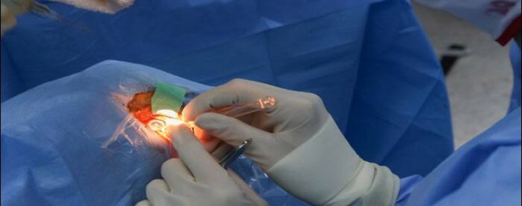
Age-related macular degeneration (AMD), a complex disease that presents pathological changes in several cell layers of the retina, including the portion known as the macula, is also a disease whose causes at the cellular level can be investigated with a careful study of dark adaptation.
Indeed, many studies showed an increase in the time taken by the rods of patients with AMD to respond to changes in illumination. This delay in rod-mediated dark adaptation in AMD has been shown to have a severity that increases with disease.
As AMD is among the leading causes of blindness in industrialised countries and no permanently effective therapy is yet available, it remains a major challenge for scientists to date.
In this context, the study of dark adaptation, being a measure that reflects structural changes in key tissues in the pathogenesis of AMD, already at very early stages of the disease, is an interesting value for clinicians to investigate.
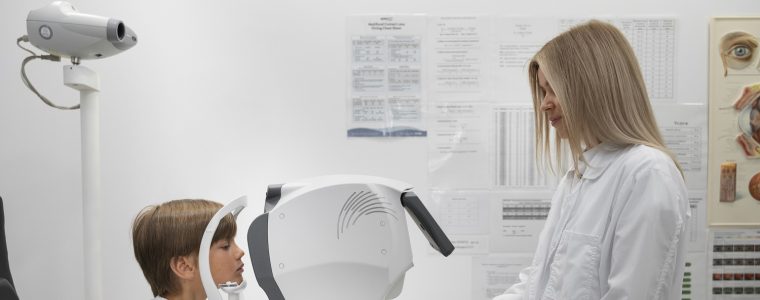
Therefore, instrumentation to test dark adaptation has seen tremendous development in recent years and, to date, several available protocols for screening AMD and monitoring disease progression use dark adaptation as an important parameter in the clinical evaluation of this maculopathy.