


Diabetic macular oedema (EMD) is one of the most serious complications of diabetic retinopathy (DR), itself a consequence of diabetic disease. Although medical progress in this area, especially in terms of treating the disease and improving visual acuity (VA), has made enormous strides, many EMD sufferers also suffer from another disorder: metamorphopsia.
What is metamorphopsia?
Metamorphopsia is a vision disorder characterised by deformed vision of images. The determining factors behind its onset are still to be clarified, just as the exact connection between metamorphopsia and diabetic macular oedema is not yet fully known.
It remains, however, certain that this visual impairment impacts negatively on quality of life of the affected person.
The Amsler test is often used to diagnose the presence of metamorphopsia in patients with various macular diseases, although the exact determination of its severity is very difficult using this test. Instead, using a more specific chart such as the M-CHARTS chart, the degrees of vertical and horizontal metamorphopsia and the degree of severity can be measured.
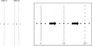
Fig. from Wada I. Quantifying metamorphopsia with M-CHARTS in patients with idiopathic macular hole. Clin Ophthalmol. 2017; 11: 1719-1726.
Metamorphopsia and Diabetic Macular Oedema
Although data in the literature to date are discordant precisely because of the lack of knowledge of the correlation mechanisms between the two diseases, some clinical evidence has shown that, through the use of M-CHARTS charts, the 46.6% of eyes with EMD presents metamorphopsia disorders. In order, therefore, to shed more light on the factors associated with the presence of metamorphopsia secondary to EMD, a study analysed 37 eyes with EMD by evaluating a number of optical coherence tomography (OCT) parameters, including disorganisation of the retinal inner layers (DRIL).
The results of the study showed that as many as 54% of the eyes examined had metamorphopsia, which was more prominent in elderly patients. Furthermore, AV was significantly better in subjects without this disorder, while DRIL was clearly higher in eyes with metamorphopsia.
The authors therefore concluded that the disorganisation of the inner retinal layers appears to play a crucial role in determining the metamorphopsia of eyes with EMD, suggesting that this visual alteration should be taken into account during the treatment of EMD.
Sources:
- Nakano E et al. Correlation between metamorphopsia and disorganization of the retinal inner layers in eyes with diabetic macular edema. Graeve's Archive for Clinical and Experimental Ophthalmology 2019; 1-6
- Wada I. Quantifying metamorphopsia with M-CHARTS in patients with idiopathic macular hole. Clin Ophthalmol. 2017; 11: 1719-1726.
Dr. Carmelo Chines
Direttore responsabile