


It is a viable option for managing rhegmatogenous retinal detachment in children.
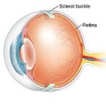
The scleral cerclage of the eye can be a major surgical technique to repair rhegmatogenous retinal detachment in paediatric patients.
As an ocular surgery technique, scleral cerclage has been successfully used to repair rhegmatogenous retinal detachments for more than 60 years. In recent decades it has been joined in the armamentarium of retinal surgeons by the pneumatic retinopexy and the vitrectomy and has experienced a partial decline in popularity, especially among patients pseudophakics.
Scleral cerclage of the eye continues, however, to be a valid procedure in countless scenarios, including paediatric eye surgery.
This specific application was the subject of a retrospective study, published in Ophthalmology, which looked at 212 eyes of children with rhegmatogenous retinal detachment who underwent reparative surgery between 2001 and 2015, with a minimum follow-up of three months. The patients were divided into three age groups: the first from 0 to 6 years, the second from 7 to 12 years and the third from 13 to 18 years.
The data report positive results overall, with a 78% of screens completely reattached in the final follow-up.
Subtotal detachments were more likely than total detachments to be successfully repaired, as opposed to the retinas of eyes that had previously undergone vitreoretinal treatment.
There were no significant variations in the success rate between the three different age groups, while they were higher in the case of primary scleral cerclage and in the case of a cerclage/vitrectomy combination via pars plan (VRS) compared to the assumption of VRS alone.
The retrospective nature of the study could be a limitation, as the minimum follow-up, adopted as an inclusion criterion, was at least three months, but some young patients were followed up for a year or even longer. However, patients with poorer eyesight or with retinal detachments recurrences might have been in the group with shorter follow-up than patients with better quality of vision and attached retinas. The different follow-up durations may, therefore, have had some effect on the accuracy of the reported findings.
Clinical relevance of scleral cerclage of the eye
Previous reviews published in the literature on rhegmatogenous retinal detachments in paediatric patients provide useful elements for evaluating their clinical and anatomical aspects, but most of these analyses are based on surgical techniques used in the era before modern vitrectomy.
Study published in Ophthalmology confirms that retinal detachment regmatogen in paediatric patients is particularly complex to treat, but that success in achieving retinal re-attachment is possible in most cases.
In this sense, primary cerclage or cerclage combined with PPV is the more effective restorative strategy than PPV alone.
All this confirms the importance of training young surgeons in the use of this surgical technique.
For this purpose, for conceptual and descriptive aspects, the tutorial prepared by the A.A.O. can be useful for teaching purposes.
Scleral buckling for rhegmatogenous retinal detachment
Sources
1. Smith JM, Ward LT, Townsend JH, et al. Rhegmatogenous Retinal Detachment in Children Clinical Factors Predictive of Successful Surgical. Ophthalmology. 2019 Sep;126(9):1263-1270.
Dr. Carmelo Chines
Direttore responsabile